
Is Ultrasound Considered a Radiology Service?
- Feb 24
- 6 min read
When you receive a letter to attend for an ultrasound scan and it states to report to the radiology department you may question: does ultrasound come under radiology? It’s a valid question. The answer is yes and no.
Let’s start with a brief explanation about where ultrasound services in London sit under the medical imaging umbrella. This will ensure you are feeling clued up before attending for your scan, whether it be a pregnancy or pelvic scan or a general check up.
What Is Radiology, Exactly?
Radiology diagnoses diseases and medical conditions using medical imaging technologies that allow doctors to view inside the human body. Radiology aids in identifying injury and illness, as well as deciding on treatment options. While radiology conjures images of X-rays for most people, it’s actually an umbrella term for all the different types of medical imaging modalities. These include X-rays, CT scans, MRI, Nuclear Medicine scans, and ultrasound.
Radiology departments at NHS hospitals throughout the UK are home to all of the above modalities. They’re nearly always called the Radiology or Medical Imaging Department and patients are normally referred there for pelvic scans, obstetric scans, and musculoskeletal scans along with CT and MRIs.
Yes, technically ultrasound falls under the category of radiology. Ultrasound is part of diagnostic imaging, the bigger branch that radiologists and sonographers both specialize in.
However, that doesn’t mean that ultrasound is the same thing as radiology. Here’s where things get a bit more, finely detailed.
How Ultrasound Differs From Other Radiology Modalities
Arguably the single biggest difference between ultrasound and most other radiology modalities is one that’s seldom talked about: ultrasound does not use radiation.
Radiation exposes patients to ionising electromagnetic waves when they undergo X-rays, CT scans and fluoroscopic examinations. Private Ultrasound scanning uses something completely different. High frequency sound waves are projected into the body. These sound waves are then reflected back by your internal tissues and organs and a computer turns those echoes into real-time visualisations on a screen.
Since ionising radiation is not involved, ultrasound exams are considered safe for every patient population including pregnant women and children, and can be repeated any number of times.
MRI scanners also don’t expose patients to radiation, instead using magnets and radio waves to create images. Both MRI and ultrasound exist within medical imaging yet function as non-ionising modalities.
Who Performs Ultrasound Scans?
At most NHS trusts, ultrasound scans are carried out by one of two types of practitioner:
A sonographer, a healthcare professional who has completed a postgraduate qualification specifically in diagnostic ultrasound. Many sonographers began their careers as radiographers or midwives before undertaking specialist ultrasound training.
A consultant radiologist is a doctor with specialist training in medical imaging who may perform more complex or interventional ultrasound procedures.
Sonographers perform ultrasound scanning as opposed to radiographers who operate radiation emitting equipment, which further demonstrates that ultrasound is its own unique specialty within radiology. Ultrasound departments in hospitals can be semi-autonomous from radiology, especially with regards to obstetric scanning where you will often find midwife sonographers.
At myGynaePlus, all of our ultrasound scans are performed by our senior consultants and sonographers who will interpret and report the findings to you on the day.
Is Ultrasound Considered a Radiology Service in the UK? The Short Answer
Yes and no, depending on context.
In the administrative and clinical framework used by the NHS and most private providers, ultrasound is classified under radiology or medical imaging. You will typically find it listed under radiology departments, and consultant radiologists oversee reporting in many settings.
Technically speaking, however, radiology in its strictest definition refers to imaging that uses radiation. Ultrasound does not. For this reason, some clinicians and professional bodies draw a distinction between radiology (radiation-based) and sonography (sound-wave-based), treating them as related but separate disciplines within the broader category of diagnostic medical imaging.
In everyday clinical practice in the UK, the terms are used interchangeably by patients and staff alike. The practical takeaway: if your GP or consultant refers you to radiology for an ultrasound, that is standard practice, and you will be seen by staff trained specifically in ultrasound scanning.
Types of Ultrasound Scans Used in Women's Health
Ultrasound is particularly well-suited to gynaecological services and obstetric care because the pelvis and abdomen respond well to sound-wave imaging, and the absence of radiation makes it appropriate at every stage of life, including pregnancy.
Here is a quick overview of the main types of ultrasound scans used in women's health:
Transabdominal ultrasound: The probe is placed on the surface of the abdomen. This is the scan most people are familiar with from pregnancy imaging. It gives a broad view of the uterus, ovaries, bladder, and surrounding structures.
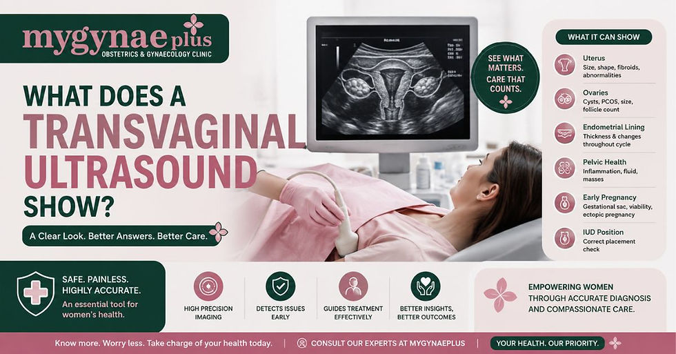
Transvaginal ultrasound: A slender probe is gently inserted into the vagina to produce closer, more detailed images of the uterus and ovaries. This is often preferred for early pregnancy scanning, follicle tracking, and assessment of conditions such as fibroids, endometriosis, or ovarian cysts, where greater image resolution is clinically useful.
Early pregnancy viability scan: Typically performed from around six weeks of gestation to confirm the pregnancy is progressing normally, check the foetal heartbeat, and establish accurate dating.
Nuchal translucency scan: Usually carried out between 11 and 14 weeks of pregnancy, this scan measures a fluid-filled space at the back of the baby's neck as part of screening for chromosomal conditions.
Anomaly scan: Performed around 20 weeks, this detailed scan checks the baby's anatomy, the position of the placenta, and the volume of amniotic fluid.
Growth and wellbeing scans: Carried out in the third trimester to monitor foetal growth, position, and movements ahead of delivery.
Follicle tracking scans Used during fertility treatment to monitor the development of follicles in the ovaries in the lead-up to ovulation or egg collection.
When Would a Doctor Refer You for a Gynaecological Ultrasound?
GPs and gynaecologists use pelvic ultrasound as a first-line investigation for a wide range of symptoms and concerns. Common reasons include:
Irregular, heavy, or painful periods
Pelvic pain or suspected endometriosis
Suspected fibroids or ovarian cysts
Postmenopausal bleeding
Difficulties conceiving or fertility investigations
Monitoring known gynaecological conditions
Confirming early pregnancy and checking viability
Ultrasound is often the starting point because it is safe, does not involve radiation, produces results in real time, and gives clinicians a clear view of soft tissue structures that X-rays cannot image well.
What Happens During a Pelvic Ultrasound?
If you have not had one before, knowing what to expect can help reduce any anxiety. Here is how a standard pelvic ultrasound typically goes:
You will be asked to lie on a couch, and gel will be applied to your lower abdomen.
The sonographer moves a handheld probe across your skin while viewing live images on a monitor.
If a transvaginal scan is needed, the sonographer will explain this fully and ask for your consent before proceeding.
The scan is generally painless, though you may feel some mild pressure.
Most pelvic scans take between 20 and 40 minutes.
Results are reviewed and a report is prepared at clinics like myGynaePlus, this happens on the same day.
You are always entitled to ask questions at any point, and you can withdraw consent for any part of the scan at any time.
Can You Self-Refer for an Ultrasound Scan?
Ultrasound scans on the NHS are normally requested by your GP or through hospital referral. Most private clinics offer a consultant led service including specialist women's health clinics who will see GP referrals and patients who refer themselves.
Self-referral means that you can book an appointment without having to see your GP beforehand. This can be helpful if you're looking for a quicker appointment or if you have a health concern that you want checked quickly. It also allows you to visit a clinic which specialises in women's health and gynaecological scanning.
FAQs
Is ultrasound a type of radiology?
Ultrasound is grouped under medical imaging and is administered within radiology departments in most UK hospitals. Technically, it is distinct from radiation-based imaging methods like X-rays and CT scans, as it uses sound waves rather than ionising radiation. Most clinicians and NHS services treat it as part of the broader radiology and imaging specialty.
Does ultrasound use radiation?
No. Ultrasound uses high-frequency sound waves, not radiation. This makes it safe for pregnant women, young children, and patients who require repeated scans. It is one of the reasons ultrasound is used routinely throughout pregnancy and for gynaecological assessments at any age.
Who carries out an ultrasound scan in the UK?
In the UK, ultrasound scans are performed by sonographers healthcare professionals with specialist postgraduate training in ultrasound or by consultant radiologists for more complex examinations. In obstetric settings, midwife sonographers are also common. All are required to meet professional standards set by regulatory bodies.
What is the difference between a sonographer and a radiologist?
A radiologist is a doctor who has completed medical training followed by specialist training in interpreting medical images across all modalities. A sonographer is a non-medical imaging professional who has trained specifically to perform and report ultrasound scans. Radiologists tend to oversee complex cases; sonographers handle the day-to-day scanning work in most departments.
Can I get a pelvic ultrasound without a GP referral?
Yes many private clinics accept self-referrals for pelvic ultrasound. You do not always need to see a GP first if you choose a private provider. This can be a quicker route if you have a specific concern, want reassurance, or are managing a known condition like endometriosis, fibroids, or PCOS and need a scan for monitoring purposes.


Comments